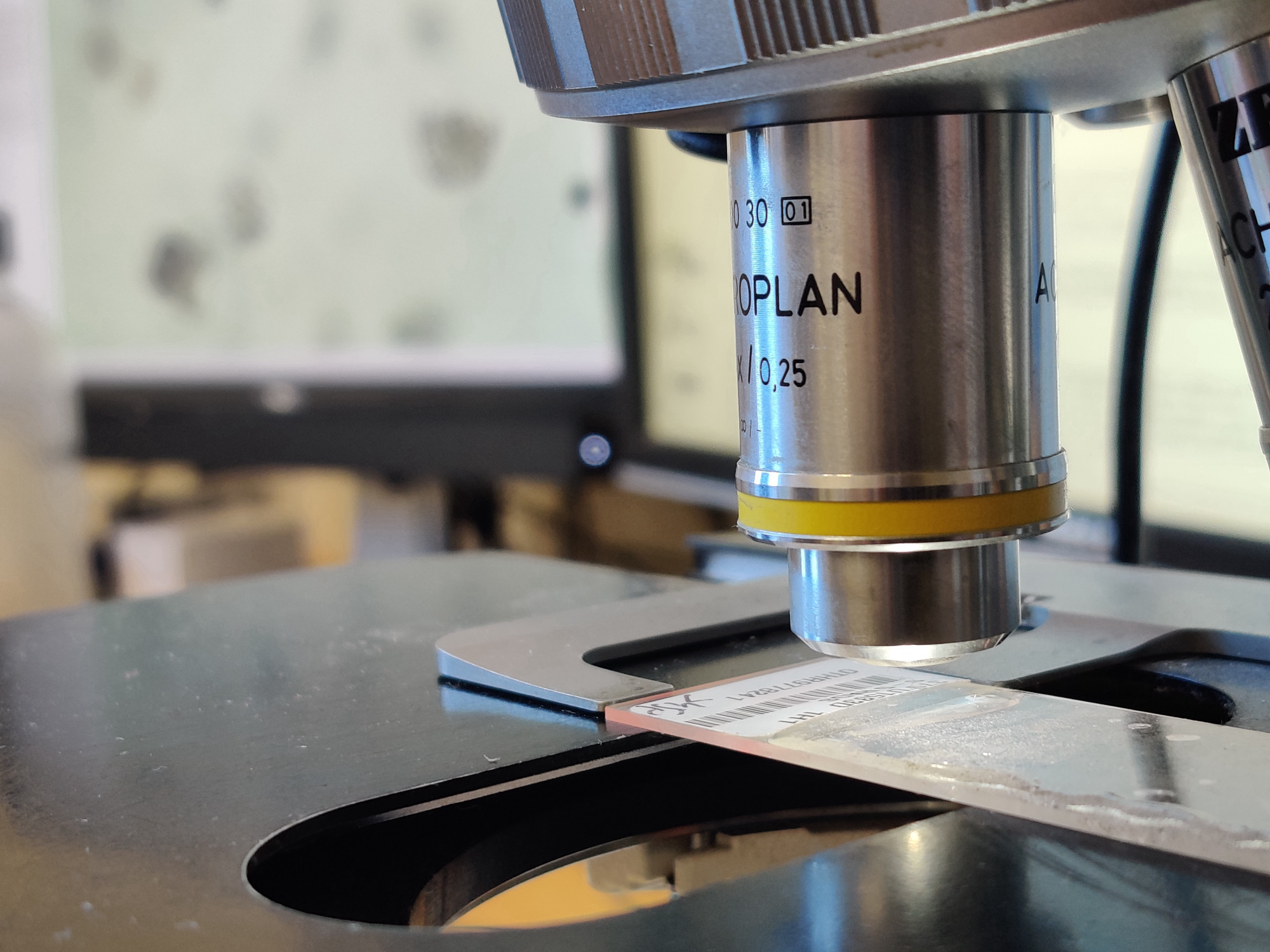
The microscopy set up on micropaleontologist Johan Renaudie’s desk in the Museum für Naturkunde Berlin, showing a radiolarian slide collected during a  deep sea drilling mission and its magnified image on the computer screen. (Image: Filippo Bertoni/MfN. All rights reserved.)
In order to explore the transformations of animals into objects that are at work in natural history and zoological collections, I zoom in on microorganisms. This is because the  microbial worlds they inhabit and shape complicate both terms of this relation in surprising ways. Microorganisms are not, technically, animals: instead they show how life forms are transformed by the categories we use to think about them, but are not bound to them and often exceed them.1 At the same time, microbial life also complicates received notions of objects as ‘things-in-themselves’, thanks to their slippery existences just outside the reach of our perception. As microorganisms are not immediately visible to the human eye, their study and understanding depends on a vast array of media and technologies to visualise them. Hence, we can never know microorganisms ‘in-themselves’, but always through specific technical set-ups. The video below illustrates this: even just four simple optical microscopy techniques bring into view four different versions of microorganisms which are incommensurable. There isn’t a version that is ‘more real’ than the others, as microorganisms exceed the limits of our perception and are only partially described by each of these techniques. This is an important reminder that how we perceive and know nature is never universal or neutral. Moreover, rather than merely describing the world, the mediations we rely on actively transform what world we know and live in. To illustrate this point better, in this story I focus on one specific microorganism,
  Cycladophora davisiana, and trace some of the mediations through which I came to know it, and how these in turn transformed and continue transforming the world. In telling this story, I focus in particular on various microscopic media that allow experts to know microbes. I start from microscopes, slides and laboratory glassware – the most obvious tools to see microorganisms. But soon less obvious tools like lists, records, publications, and digital databases used in
  recording their worlds emerge as equally crucial mediations. These bring with them also broader technical systems, like deep sea drilling,
  taxonomic orders, and natural history collections themselves – like the
  Lamont-Doherty Collection and the
  NSB Database. In this sense, the mediations involved in knowing C. davisiana and other microorganisms extend beyond the bounds of what we usually imagine as matters of perception or microscopy.2 This reminds us that the world is not closed off in a stable order that is down to us to decipher, but that ordering and sense-making are ongoing processes involved in worldly transformativity and the continuous becoming we are also part of, together with countless others. And that our attempts at knowing nature are always situated and partial, but also exceed a simplistic view of self-contained subjects knowing a world of equally bounded objects – too often still implicit in Western notions of knowledge and vision. Objectivities are hard-won and fragile achievements, that require lots of work from many different people, objects, and entities and rely on a multiplicity of on-going mediations: keeping this in mind is crucial to better grasp the role of natural history collections, and their importance in
  shaping the world we live in – and hopefully can help in making them more just and critical in the future.
This episode of the beautiful webseries “Journey to the Microcosmos” illustrates four different techniques for optical microscopy. The resulting images of the microscopic world remind us of the importance of media in studying microorganisms and the world at large. What we see changes how we see. (Source: Journey to the Microcosmos/YouTube)
As I recount in the story about  finding Cycladophora, the first specimen of this organism to be described was hauled from the bottom of Davis Strait, off the coast of Greenland, in 1859 – as part of a survey for a never-completed transatlantic telegraph line. The sediment samples collected in this expedition were sent to Berlin to be analysed by Christian Gottfried Ehrenberg, an authority on
  Infusoria – as microbes were known at the time. In what was to become the Berlin Natural History Museum, the microorganisms contained in the samples went through important transformations, which allowed Ehrenberg to
  classify Cycladophora. Crucial to this classification and inscription of Cycladophora davisiana into taxonomic orders, were Ehrenberg’s microscopic observations. Placing the sieved and washed samples on a transparent mica disk under an optical microscope – an apparatus consisting of lenses that allow to magnify small preparations by diffracting and manipulating light3 – Ehrenberg could observe the intricate details of the microscopic remains of this microorganism, describe them, and classify C. davisiana as a
  Radiolaria. Because of the centrality of this mediation, the history of the study of microorganisms has been told many times as a progression of technological innovations in the field of microscopy.4 While the optical microscope and its descendants are central in these mediations, as historians of science have shown they also depend on a number of other, more mundane and yet far-reaching tools, techniques, and practices: the definition and standardisation of optics and resolution powers,5 as well as of units of measure, are central examples here, as are the development of standardised laboratory glassware,6 like microscope slides and Petri dishes,7 but also the use of
  labels,
  lists, illustrations,8 photographs, videos,9 and other documents to record the diversity of nature, and the parallel development of accounting practices and the political economies of collecting. In the study of living microorganisms, these developments also entwined with the media for growing them in the lab (like the most common medium for microbial cultures, agar10), and the complex culturing techniques that continue to allow scientists to grow microbes under controlled conditions.11
While benefiting from the lessons learned through these techniques, micropaleontology mostly deals with long dead microfossils, which are more stable and easier to study, manipulate, and preserve.12 So, even if C. davisiana still resists culturing in the lab to this day, the study of its fossilised remains bypasses the fragile living forms and concentrates instead on the morphology and physicochemical characteristics of this organism’s durable microscopic shell, known as test. It was this feature that allowed the early classification of Cycladophora in taxonomic orders, and engendered a number of ways of  using Cylcladophora that are central to how this organism is currently understood and in turned shape the world it inhabits. This is evident in the central use of microfossils like C. davisiana in
  biostratigraphy: by indexing geological and planetary changes, its fossilised tests are mobilised to unfold
  micropaleontological formations. These micropaleontological studies allow scientists to trace the biogeochemical connections between
  microbes and the planet: the accumulation and charting of data, specimens, and
  core samples laid the foundations for present-day models of planetary dynamics and continue to inform how the planet is treated. Simultaneously, through the development of
  industrial micropaleontology, microfossils also play a crucial role in the continued extraction and exploitation of
  fossil fuels, which in turn is affecting global climate in ways that impact all of us. In this sense, the mediations that make microscopic worlds visible reach much further than the objectives and oculars of microscopes, ranging from the molecular intricacies of physicochemical matters, on to the planetary vision assembled by charting global distributions and changes in microfossil formations. As they make these otherwise incommensurable scales easier to handle, the transformations of animals into objects traced in this website also make evident that how we know the world also actively affects what worlds come to matter, and for whom. Rather than self-contained and autonomous ‘things-in-themselves’, the objects that C. davisiana is transformed into are situated and temporary achievements that require complex relational efforts to take and maintain specific forms. It is this relationality, and the countless mediations that it involves, that remind us that how we see nature also reciprocally has a part in shaping what natures come to take form. Like the microscope, natural history collections like those of the Museum für Naturkunde Berlin, then, transform invisible microbial life forms into scientific objects and data, not only making them visible, but also bringing them to matter far beyond their limited ranges – as they come to shape the planet itself.
- For an overview of how contemporary biology is challenging traditional notions of bounded individuals, importantly influenced by microbial sciences, read Scott F. Gilbert, J. Sapp, and A.I. Tauber. “A Symbiotic View of Life: We Have Never Been Individuals”. The Quarterly Review of Biology 87, no. 4 (2012): 325-41. http://doi:10.1086/668166.↩
- To better understand the complex implications of the sensorial and perception in the work of science, you can read this blog post I wrote: Society for Cultural Anthropology. https://culanth.org/fieldsights/science-and-the-senses-perturbation (17.01.2022).↩
- Selectively allowing to manipulate light, glass has been key to peeking into the microcosmos: the optical properties of glass are central to all these distributed technical apparatuses of sense-making. Interestingly, they are also responsible for the diaphanous and beautiful structures that constitute the translucent microscopic shells of many microorganisms, including radiolarians and foraminiferans. In fact, glass is made out of sand, which is formed by the sedimentation and weathering of large masses of microfossils over geological times. It is the very bodies of microorganisms like Cycladophora that become the material medium that makes microscopic observations possible.↩
- For an overview of this history of microscopy, you can visit: Britannica Online. https://www.britannica.com/technology/microscope/History-of-optical-microscopes (17.01.2022). For more resources you can also visit the website of the National Museums Scotland: https://www.nms.ac.uk/explore-our-collections/stories/science-and-technology/microscopes/ (17.01.2022). For a more in-depth historical exploration of the early phases of microscopy, see Marc J. Ratcliff. The Quest for the Invisible: Microscopy in the Enlightenment. Farnham: Ashgate Pub. Limited, 2009. For an interesting experiment in ‘predecessor sciences’ (the speculative exploration of how science could have been otherwise in the past), check out this box microscope prototype: Unterbahn.com. https://unterbahn.com/2020/12/05/box-microscopes/ (17.01.2022). Inspired by the few remaining sources about early microscopic tools beyond Eurocentric historiography, this tool was created by Jeffrey Yoo Warren, and it reminds us that when we tell the history of microscopy, we are always telling a partial and biased history that relies on situated and limited archives.↩
- The specificities of the historical standardisation of microscope resolution, along with the convergence of microscopy and life sciences in 19th century Germany, are treated in detail in Jutta Schickore. The Microscope and the Eye: A History of Reflections, 1740-1870. Chicago: University of Chicago Press, 2007.↩
- To learn more about the important role of standardised glassware in the life sciences, you can read this fascinating article: Kijan Espahangizi. “The Twofold History of Laboratory Glassware”. In Membranes, Surfaces, Boundaries: Interstices in the History of Science, Technology and Culture. Mathias Grote, Max Stadler und Laura Otis (Hg.). Berlin: Max-Planck-Inst. für Wissenschaftsgeschichte, 2011: 17-33.↩
- To learn more about the ubiquitous Petri dish, visit, The Biomedical Scientist. https://thebiomedicalscientist.net/science/big-story-petri-dish or, Somatosphere.net. http://somatosphere.net/2014/petri-dish.html/ (17.01.2022). To better understand its important role in consolidating a specific version of microbiology at the expense of alternative ones, you can read this article by geographer Frederick Attenborough. “The Monad and the Nomad: Medical Microbiology and the Politics and Possibilities of the Mobile Microbe”. Cultural Geographies 18, no. 1 (2011): 91-114.↩
- For an impressive example of the importance and beauty of these early illustrations, have a look at Hooke’s famous 1665 book Micrographia and its captivating plates, which can be browsed digitally here: The Royal Society. https://royalsociety.org/blog/2020/07/micrographia-online/. Also, check out this episode of “Journey to the Microcosmos” on Ehrenberg’s own illustrations held at the Museum für Naturkunde Berlin: YouTube. https://youtu.be/PKMUJdn09OU (17.01.2022).↩
- See, for instance, these articles: Hannah Landecker. “Microcinematography and the History of Science and Film”. Isis 97, no. 1 (2006): 121-132. https://doi.org/10.1086/501105; and Hannah Landecker. “Seeing Things: From Microcinematography to Live Cell Imaging”. Nature Methods 6, no. 10 (2009): 707-709. https://doi.org/10.1038/nmeth1009-707.↩
- You can read the fascinating story of Angelina Fanny Hesse’s central role in the key innovation represented by the agar as a medium for cell culture here: Popular Science. https://www.popsci.com/blog-network/ladybits/forgotten-woman-who-made-microbiology-possible/ (17.01.2022).↩
- To learn more on the important history of cell cultures, read the great book by Hannah Landecker: Culturing Life: How Cells Became Technologies. Cambride: Harvard University Press, 2007.↩
- Although even these organisms present their own challenges; you can learn more about this by reading Brian Bracegirdle. “A History of Microtechnique”. Journal of the History of Biology 14, no. 2 (1981). For the more technical side of this, have a look at: Birger Neuhaus, Thomas Schmid, and Jens Riedel. “Collection Management and Study of Microscope Slides: Storage, Profiling, Deterioration, Restoration Procedures, and General Recommendations”. Zootaxa 4322, no. 1 (2017): 1-173.↩