Papier mâché Bombyx mori by Louis Auzoux in the Zoological Teaching Collection of the Humboldt-Universität zu Berlin. (Image: Kerstin Stoll/Zoological Teaching Collection. All rights reserved.)
In the 19th and 20th centuries, a wide range of materials were used to make both scientific and mainstream animal models – long before  plastic models were commonly used. The selection of materials influenced not just their malleability but also their potential colouring, weight, and durability, as well as how many copies could be produced and how they would be circulated. These factors therefore had an impact on practical questions of utilisation, on aesthetic issues, and on the knowledge that a model should or could convey.
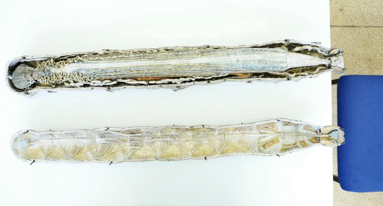
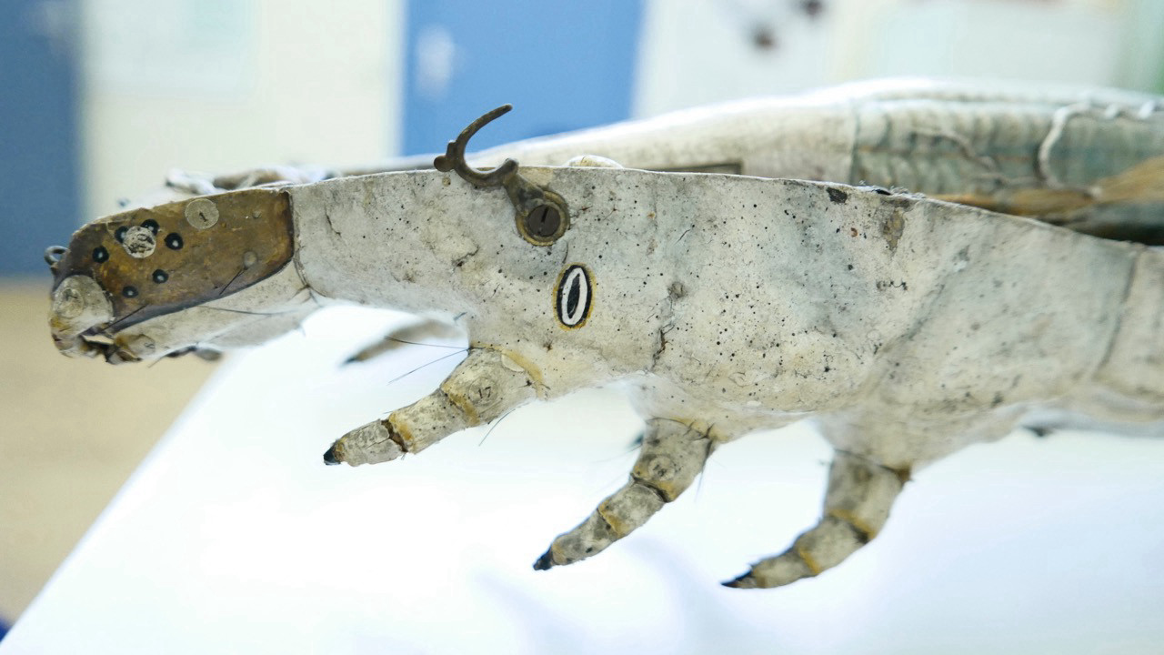
While the studio of Adolf Ziegler (1820-1889) and his son Friedrich (1860-1936) produced models of animal and human anatomy in wax1 in order to emphasise their translucent fleshiness, the anatomist Louis Auzoux (1792-1880) chose papier mâché to make his animal and plant models.2 This technique had already been utilised in China to make helmets and in Europe at the beginning of the 19th century to construct anatomical models, but Auzoux developed a new formula, which he carefully guarded. The lightness of the dried mass made from paper, various other ingredients, and adhesive agents allowed, for example, for a small, approximately five-centimetre-long silkworm to be enlarged to a length of 75 centimetres and for its body parts to be presented naturalistically.3
Even with the lid closed, the insides of the silkworm can be seen in the papier mâché model. (Image: Kerstin Stoll/Zoological Teaching Collection. All rights reserved.)
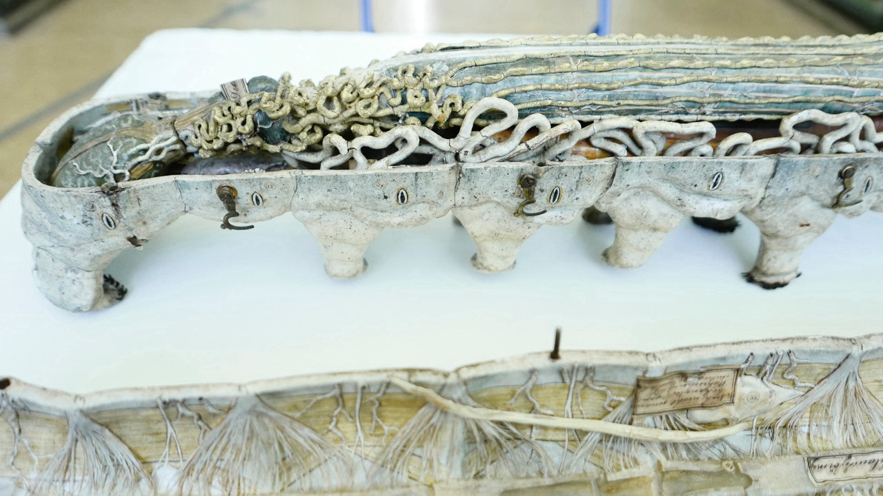
The functional, collapsible anatomical model (which Auzoux also referred to as an ‘anatomie clastique’) of the Bombyx mori, the  silkworm, which used to be bred for
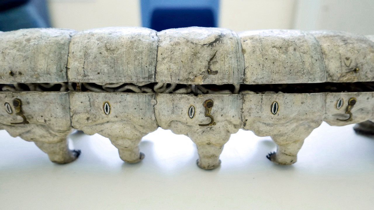
  cultivating silk in Berlin and Brandenburg, shows its skin and extremities, its inner organs, and its silk glands considerably enlarged and in great detail. This carefully painted artefact, which was probably produced in the 1860s and is still housed in the
  Zoological Teaching Collection of the Humboldt-Universität zu Berlin,4 can be opened or closed using metal hooks and, alongside some numbering, also
  displays text that has been written directly onto the individual body parts. This writing does not just make reference to the body parts like a legend; rather, it is inscribed onto the model in ink and letters and simultaneously describes it in scientific jargon. The silkworm appears here as a ‘working animal’ that turns out a product – silk thread. At the same time, the model represents one stage of a complete metamorphosis, whose imago (the sexually mature insect after its final transformation) Auzoux has immortalised in papier mâché. He also had models of female and male silk moths on offer for teaching purposes.
- Cf. Nick Hopwood. Embryos in Wax: Models from the Ziegler Studio. Cambridge: Whipple Museum of the History of Science, 2002.↩
- On Auzoux’ models, see Bart Grob. The World of Auzoux: Models of Man and Beast in Papier-Mâché. Leiden: Museum Boerhaave, 2000; Margret Maria Cocks. “Dr Louis Auzoux and His Collection of Papier-Mâché Flowers, Fruits and Seeds”. Journal of the History of Collections 26, no. 2 (2014): 229-248; Margret Maria Olszewski. “Dr. Auzoux’ Botanical Teaching Models and Medical Education at the Universities of Glasgow and Aberdeen”. Studies in History and Philosophy of Science. Part C 42, no. 3 (2011): 285-296.↩
- Cf. “Modell, Raupe, Seidenspinner”. Database entry of the Humboldt-Universität zu Berlin, no date, https://www.sammlungen.hu-berlin.de/objekte/zoologische-lehrsammlung/8322/ (03.01.2022).↩
- Cf. Dr. L. Wittmack. Allgemeiner Katalog des königlichen landwirthschaftlichen Museums zu Berlin, Berlin: Verlagsbuchhandlung Wiegandt und Hempel, 1869: 108-109. The model was purchased from Auzoux for 66 talers, and it is highly likely that it then moved to Invalidenstraße 42 when the United Agricultural Teaching Institute and Museum (Vereinigtes landwirthschaftliches Lehr-Institut und Museum) was built in 1880.↩